How many cavities is the heart divided
How many cavities is the heart divided
The Anatomy of the Heart, Its Structures, and Functions
The heart is the organ that helps supply blood and oxygen to all parts of the body. It is divided by a partition (or septum) into two halves. The halves are, in turn, divided into four chambers. The heart is situated within the chest cavity and surrounded by a fluid-filled sac called the pericardium. This amazing muscle produces electrical impulses that cause the heart to contract, pumping blood throughout the body. The heart and the circulatory system together form the cardiovascular system.
Heart Anatomy
The heart is made up of four chambers:
Heart Wall
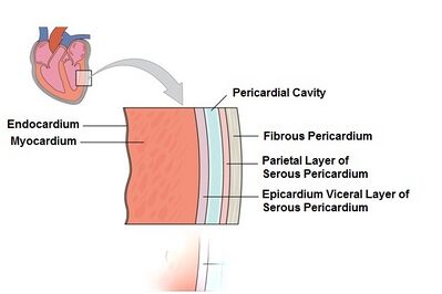
The heart wall consists of three layers:
Cardiac Conduction
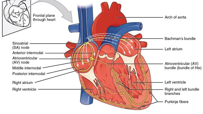
Cardiac conduction is the rate at which the heart conducts electrical impulses. Heart nodes and nerve fibers play an important role in causing the heart to contract.
Cardiac Cycle
The Cardiac cycle is the sequence of events that occurs when the heart beats. Below are the two phases of the cardiac cycle:
Valves
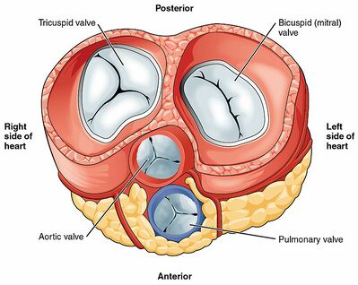
Heart valves are flap-like structures that allow blood to flow in one direction. Below are the four valves of the heart:
Blood Vessels
Blood vessels are intricate networks of hollow tubes that transport blood throughout the entire body. The following are some of the blood vessels associated with the heart:
Heart
Overview
What is the heart?
The heart is a fist-sized organ that pumps blood throughout your body. It’s the primary organ of your circulatory system.
Your heart contains four main sections (chambers) made of muscle and powered by electrical impulses. Your brain and nervous system direct your heart’s function.
What does a heart diagram look like?
The inside and outside of your heart contain components that direct blood flow:
Inside of the Heart
Outside of the Heart
Function
What is the heart’s function?
Your heart’s main function is to move blood throughout your body. Your heart also:
How does your heart work with other organs?
Your heart works with other body systems to control your heart rate and other body functions. The primary systems are:
Anatomy
Where is your heart located?
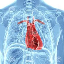
Your heart is located in the front of your chest. It sits slightly behind and to the left of your sternum (breastbone). Your ribcage protects your heart.
What side is your heart on?
Your heart is slightly on the left side of your body. It sits between your right and left lungs. The left lung is slightly smaller to make room for the heart in your left chest.
How big is your heart?
Everyone’s heart is a slightly different size. Generally, adult hearts are about the same size as two clenched fists, and children’s hearts are about the same size as one clenched fist.
How much does your heart weigh?
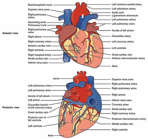
What are the parts of the heart’s anatomy?
The parts of your heart are like the parts of a house. Your heart has:
Heart walls
Your heart walls are the muscles that contract (squeeze) and relax to send blood throughout your body. A layer of muscular tissue called the septum divides your heart walls into the left and right sides.
Your heart walls have three layers:
The epicardium is one layer of your pericardium. The pericardium is a protective sac that covers your entire heart. It produces fluid to lubricate your heart and keep it from rubbing against other organs.
Heart chambers
Your heart is divided into four chambers. You have two chambers on the top (atrium, plural atria) and two on the bottom (ventricles), one on each side of the heart.
Heart valves
Your heart valves are like doors between your heart chambers. They open and close to allow blood to flow through.
The atrioventricular (AV) valves open between your upper and lower heart chambers. They include:
Semilunar (SL) valves open when blood flows out of your ventricles. They include:
Blood vessels
Your heart pumps blood through three types of blood vessels:
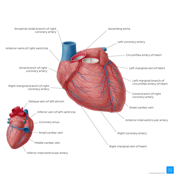
Your heart receives nutrients through a network of coronary arteries. These arteries run along your heart’s surface. They serve the heart itself.
Electrical conduction system
Your heart’s conduction system is like the electrical wiring of a house. It controls the rhythm and pace of your heartbeat. It includes:
Your heart also has a network of electrical bundles and fibers. This network includes:
Conditions and Disorders
What conditions and disorders affect the human heart?
Heart conditions are among the most common types of disorders affecting people. In the United States, heart disease is the leading cause of death for people of all genders and most ethnic and racial groups.
Common conditions that affect your heart include:
How can I keep my heart healthy?
If you have a condition that affects your heart, follow your healthcare provider’s treatment plan. It’s important to take medications as prescribed.
You can also make lifestyle changes to keep your heart healthy. You may:
Frequently Asked Questions
What should I ask my doctor about my heart?
You may want to ask your healthcare provider:
A note from Cleveland Clinic
Your heart is the primary organ of your circulatory system. It pumps blood throughout your body, controls your heart rate and maintains blood pressure. Your heart is a bit like a house. It has walls, rooms, doors, plumbing and an electrical system. All the parts of your heart work together to keep blood flowing and send nutrients to your other organs. Conditions that affect your heart are some of the most common types of conditions. Ask your healthcare provider how you can improve your heart health.
Last reviewed by a Cleveland Clinic medical professional on 08/26/2021.
References
Cleveland Clinic is a non-profit academic medical center. Advertising on our site helps support our mission. We do not endorse non-Cleveland Clinic products or services. Policy
Anatomy of the Human Heart
Contents
Introduction [ edit | edit source ]
The heart is a muscular organ that serves to collect deoxygenated blood from all parts of the body, carries it to the lungs to be oxygenated and release carbon dioxide. Then, it transports the oxygenated blood from the lungs and distributes it to all the body parts [1]
This 10 minute video is a good summary of the heart. [3]
Anatomy [ edit | edit source ]
The heart has a somewhat conical form and is enclosed by the pericardium. It is positioned posteriorly to the body of the sternum with one-third situated on the right and two-thirds on the left of the midline. The heart measures 12 x 8.5 x 6 cm and weighs
310 g (males) and
Layers of the Heart Walls [ edit | edit source ]
The heart wall consists of three layers enclosed in the pericardium [1] [5] [6] :
The rest of the heart is composed mainly of the subepicardial and subendocardial layers.
Structure and Function [ edit | edit source ]
The heart is subdivided by septa into right and left halves, and a constriction subdivides each half of the organ into two cavities, the upper cavity being called the atrium, the lower the ventricle. The heart, therefore, consists of four chambers:
It is best to remember the four chambers and four valves in order of the series that blood travels through the heart:
Heart Valves [ edit | edit source ]
The heart has four valves. All four valves of the heart have a singular purpose: allowing forward flow of blood but preventing backward flow. [7] The outflow of each chamber is guarded by a heart valve:
Atrioventricular valves between the atria and ventricles
Semilunar valves which are located in the outflow tracts of the ventricles
Blood Supply [ edit | edit source ]
The heart is supplied by two coronary arteries:
2. Right coronary artery: branches supply the right ventricle, right atrium, and left ventricle’s inferior wall.
Coronary arteries and veins course over the surface of the heart. Most coronary veins coalesce into the coronary sinus that runs in the left posterior atrioventricular groove and opens into the right atrium. Other small veins, called thebesian veins, open directly into all four chambers of the heart. [7]
Venous drainage and Lymphatics [ edit | edit source ]
The lymphatic vessels drain mainly into:
Nerve Supply [ edit | edit source ]
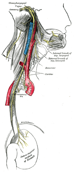
The main control of the heart resides with the medulla oblongata. There is an area called the cardioacceleratory centre, or pressor centre, in the upper part of the medulla oblongata, and an area called the cardioinhibitory centre, or depressor centre, in the lower part. Together they are called the cardioregulatory centre, since they interact to control heart rate, etc.
The nervous supply to the heart is autonomic, consisting of both sympathetic and parasympathetic parts. The sympathetic fibres arise from the pressor centre, while the parasympathetic fibres arise in the depressor centre. See also Vagal Tone
Heart Conduction System [ edit | edit source ]
An electrical conduction system regulates the pumping of the heart and timing of contraction of various chambers. Heart muscle contracts in response to the electrical stimulus received system generates electrical impulses and conducts them throughout the muscle of the heart, stimulating the heart to contract and pump blood. Among the major elements in the cardiac conduction system are the sinus node, atrioventricular node, and the autonomic nervous system.
Relevance to Physiotherapy [ edit | edit source ]
Videos [ edit | edit source ]
Heart
Author: Jana Vasković MD • Reviewer: Alexandra Osika
Last reviewed: July 06, 2022
Reading time: 12 minutes
The heart is a muscular organ that pumps blood around the body by circulating it through the circulatory/vascular system. It is found in the middle mediastinum, wrapped in a two-layered serous sac called the pericardium. The heart is shaped as a quadrangular pyramid, and orientated as if the pyramid has fallen onto one of its sides so that its base faces the posterior thoracic wall, and its apex is pointed toward the anterior thoracic wall. The great vessels that originate from the heart, radiate their branches to the head and neck, the thorax and abdomen and the upper and lower limbs.
The heart holds a special position in anatomical sciences. For instance, you can live without your spleen or with only one kidney, you can even regrow your liver–but you cannot live without a heart. This page will introduce you to the anatomy of the heart.
Heart anatomy
The heart has five surfaces: base (posterior), diaphragmatic (inferior), sternocostal (anterior), and left and right pulmonary surfaces. It also has several margins: right, left, superior, and inferior:
Inside, the heart is divided into four heart chambers: two atria (right and left) and two ventricles (right and left).
The right atrium and ventricle receive deoxygenated blood from systemic veins and pump it to the lungs, while the left atrium and ventricle receive oxygenated blood from the lungs and pump it to the systemic vessels which distribute it throughout the body.
The left and right sides of the heart are separated by the interatrial and interventricular septa which are continuous with each other. Furthermore, the atria are separated from the ventricles by the atrioventricular septa. Blood flows from the atria into the ventricles through the atrioventricular orifices (right and left)–openings in the atrioventricular septa. These openings are periodically shut and open by the heart valves, depending on the phase of the heart cycle.
Although there are a lot of structures in the heart diagrams, you shall not worry, we’ve got them all covered for you in these articles and video tutorials. Be sure to check out our specially designed heart anatomy quiz which will help you to master the heart anatomy.
Heart valves
Heart valves separate atria from ventricles, and ventricles from great vessels. The valves incorporate two or three leaflets (cusps) around the atrioventricular orifices and the roots of great vessels.
The cusps are pushed open to allow blood flow in one direction, and then closed to seal the orifices and prevent the backflow of blood. Backward prolapse of the cusps is prevented by the chordae tendineae–also known as the heart strings–fibrous cords that connect the papillary muscles of the ventricular wall to the atrioventricular valves.
There are two sets of valves: atrioventricular and semilunar. The atrioventricular valves prevent backflow from the ventricles to the atria:
It is very easy to remember which is which if you use a mnemonic! Just memorise LAB RAT and you’re set!
Left Atrium: Bicuspid
Right Atrium: Tricuspid
Semilunar valves prevent backflow from the great vessels to the ventricles.
In clinical practice, the heart valves can be auscultated, usually by using a stethoscope. In order to be successful, one needs to master the surface projections of the heart.
We have created a quiz, so you can test your newly acquired knowledge on the heart valves.
Heart valves mnemonic
Need an easy way to remember the four heart valves? Memorise the phrase ‘Try Pulling My Aorta’, which stands for:
Tricuspid
Pulmonary
Mitral
Aortic
You can then go on to solidify your knowledge about heart valve anatomy with this study unit:
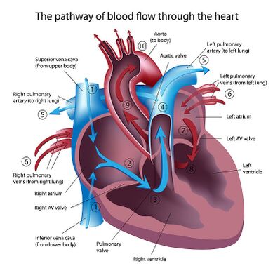
Blood flow through the heart
The blood flow through the heart is quite logical. It happens with the heart cycle, which consists of the periodical contraction and relaxation of the atrial and ventricular myocardium (heart muscle tissue). Systole is the period of contraction of the ventricular walls, while the period of ventricular relaxation is known as diastole. Note that whenever the atria contract, the ventricles are relaxed and vice versa. Let’s put into words the heart blood flow diagram:
Heart
Heart Definition
The heart is a muscular organ that pumps blood throughout the body. It is located in the middle cavity of the chest, between the lungs. In most people, the heart is located on the left side of the chest, beneath the breastbone.
The heart is composed of smooth muscle. It has four chambers which contract in a specific order, allowing the human heart to pump blood from the body to the lungs and back again with high efficiency. The heart also contains “pacemaker” cells which fire nerve impulses at regular intervals, prompting the heart muscle to contract.
This animation shows the functioning of this extraordinarily complex pump in action. As you read this article, try scrolling back up and seeing if you can spot the chambers, valves, and blood vessels we’re discussing in action:

The heart is one of the most vital and delicate organs in the body. If it does not function properly, all other organs – including the brain – begin to die from lack of oxygen within just a few minutes. As of 2009, the most common cause of death in the world was heart disease.
Most heart disease occurs as a result of age or lifestyle. Cholesterol can build up in the arteries as a person gets older, and this is more likely for people who have diets high in saturated fat and cholesterol. Rarely, however, heart disease can also occur due to a virus or bacterium that infects the heart or its protective tissues.
Scientists have had some success replicating the heart’s pumping action with artificial pumps, but these pumps can be rejected by the body, and they break down over time.
The four-chambered heart found in mammals and birds is more efficient than the one, two, or three-chambered hearts found in some other animals. It is thought that warm-blooded animals need highly efficient circulation to satisfy their cells’ high demand for oxygen. This is especially true of humans, whose huge brains require a near-constant supply of oxygen to function!
Function of the Heart
The heart pumps blood through our immense and complicated circulatory systems at high pressure. It is a truly impressive feat of engineering, as it must circulate about five liters of blood through a full 1,000 miles worth of blood vessels each minute! We will talk more about how the heart accomplishes this remarkable task under the “Heart Structure” section below.
The pumping action of the heart allows the movement of many substances between organs in the body, including nutrients, waste products, and hormones and other chemical messengers. However, arguably the most important substance it circulates is oxygen.
Oxygen is required for animal cells to perform cellular respiration. Without oxygen, cells cannot break down food to produce ATP, the cellular currency of energy. Soon, none of their energy-dependent processes can function. Without its energy-dependent processes, a cell dies.
Neural tissues, including the brain, are particularly sensitive to oxygen deprivation. Neural tissues maintain a special cellular chemistry which must be constantly maintained through the consumption of lots and lots of energy. If ATP production stops, neural cells can begin to die within minutes.
For this reason, the body has taken many special measures to protect the heart. It is located below the strongest part of the ribcage and cushioned between the lungs. It is also surrounded by a protective membrane called the pericardium, which is filled with additional cushioning fluid.
Heart Structure
The heart’s unique design allows it to accomplish the incredible task of circulating blood through the human body. Here we will review its essential components, and how and why blood passes through them.
Layers of the Heart Wall
The heart has three layers of tissue, each of which serve a slightly different purpose. These are:
Chambers of the Heart
The heart has four chambers, which are designed to pump blood from the body to the lungs and back again with extremely high efficiency. Here we’ll see what the four chambers are, and how they do their jobs:
After the blood has circulated through the body and the oxygen has been exchange for carbon dioxide waste from the body’s cells, the blood re-enters the right atrium and the process begins again.
In most people, this whole circulatory path only takes about a minute to complete!
Valves of the Heart
You may be wondering how the heart ensures that blood flows in the right direction between these chambers and blood vessels. You may also have heard of “heart valves” referred to in a medical context.
Heart valves are just that – biological valves that only allow blood to flow through the heart in one direction, ensuring that all the blood gets to where it needs to be.
Here is a list of the most important valves in the heart, and an explanation of why they are important:
Many people have minor irregularities with these valves, such as mitral valve prolapse, which make their hearts less efficient or more prone to experiencing problems. People with minor valve issues can often lead a normal, healthy life.
However, total failure of any of these valves can be catastrophic for the heart and for blood flow. That’s why people with conditions like mitral valve prolapse are often turned down by the military and other programs that involve conditions which can be very taxing for the heart.
The Sinoatrial Node
The sinoatrial node is another very important part of the heart. It is a group of cells in the wall of the right atrium of the heart – and it is what keeps the heart pumping!
The cells in the sinoatrial node produce small electrical impulses in a regular rhythm. These impulses are what drive the contractions of the four chambers of the heart.
Artificial pacemakers replicate the action of the sinoatrial node by making similar electrical impulses for people whose sinoatrial node isn’t functioning properly. However, healthy people have a natural pacemaker built right into the heart!
1. Which of the following is NOT true of the human heart?
A. It has four chambers.
B. It has a built-in “pacemaker” called the sinoatrial node.
C. It is responsible for pumping oxygen-filled blood around the body.
D. It is the only organ in the body you cannot live without.
2. Which of the following is NOT a layer of the heart?
A. The myocardium
B. The endocardium
C. The myometrium
D. The epicardium
3. Why does the heart need valves?
A. To keep out pathogens and toxins that could damage the heart
B. To ensure that blood goes not flow backward into the wrong chamber of the heart
C. To ensure that the heart does not pump too much blood at once
D. None of the above
:max_bytes(150000):strip_icc()/heart-2607178_1920-9b2a3115e5ea43f5b3f50af576c4a524.jpg)

:background_color(FFFFFF):format(jpeg)