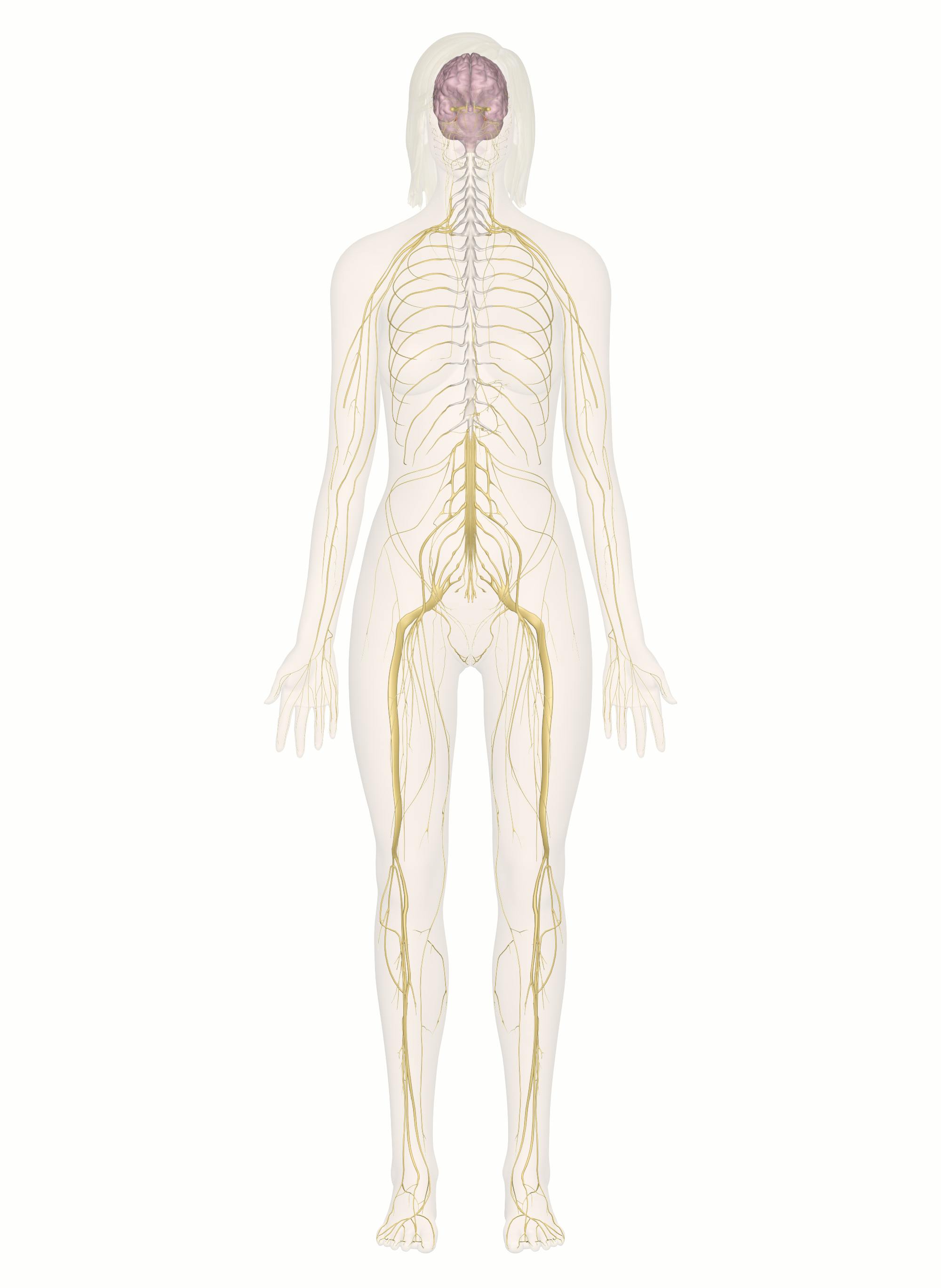
What organs are made up nervous system
What organs are made up nervous system
The Nervous System
Hey, guys! Welcome to this Mometrix video over the nervous system.
All systems and parts of the body are important, but the nervous system is the system that has its fingers in everything your body does. It is the system that literally controls every process within your body.
The nervous system includes anything within the body that would allow you to sense; so, the brain, spine and spinal cord, all nerves, and any other sensory neurons.
Nervous System Function
The nervous system has four main roles or functions:
First, to collect and receive information internally and externally; so, from within the body and outside of the body.
Second, to process or derive meaning from the information collected and received.
Third, after the information has been processed, the nervous system forwards that information to the appropriate party; so, to the muscles, organs, or glands. This information sets them up to react how they need to.
Fourth, the nervous system dispatches the information to the regions of the brain that handle the processing of the information.
The nervous system, just like every other structure and makeup of our bodies, is very ordered and systematic. You can think of the nervous system as having two main divisions, and then several subdivisions that fit within those two divisions. The two divisions of the nervous system are the central nervous system (CNS) and the peripheral nervous system (PNS).
First, we will take a look at the central nervous system, and what exactly that includes.
Central Nervous System Function
The main function of the central nervous system is to incorporate the information it is getting and systemize the information so that it can queue a proper response.
The central nervous system is composed of the brain and the spinal cord. These two components encompass several other components. So, we will take a closer look at the brain first.
Regions of the Brain
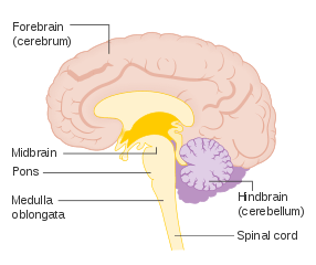
The brain has six different regions: the cerebellum, cerebrum, medulla, brainstem, thalamus, and hypothalamus.
Cerebellum
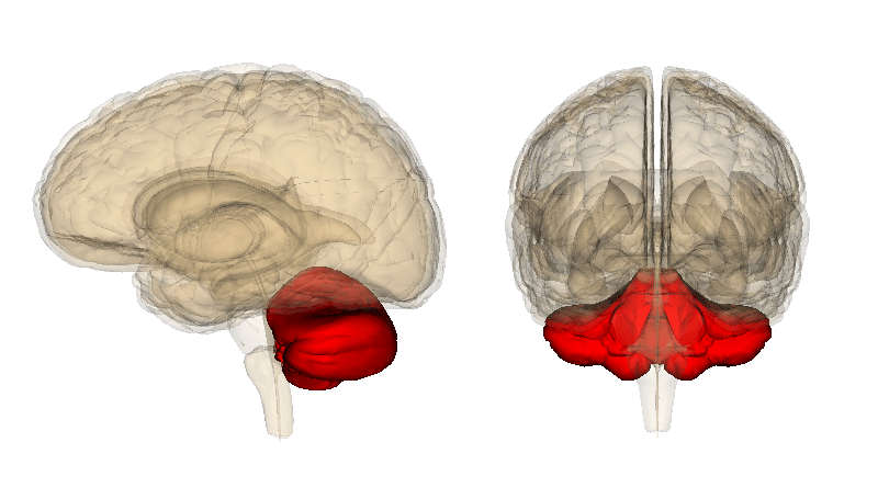
The cerebellum sits right behind the upper portion of the brain stem (which is where the brain and spinal cord connect) and consists of two half-spheres (or hemispheres).
The cerebellum is the region of the brain that coordinates movement, and where various features of motor learning take place. In other words, the cerebellum is the part of your brain that allows for balance, coordination, speech, and posture, as well as things like muscle memory.
Cerebrum
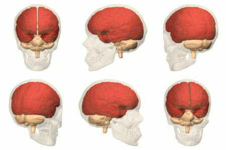
The cerebrum sits in the top portion of the cranial cavity. The cerebrum is separated into a left and right hemisphere by a trench-like line called the longitudinal fissure. Now, perhaps counterintuitively, the left half of the cerebrum manages the right side of the body and the right half manages the left side of the body. However, even though there is a groove that distinctly separates the two halves of the cerebrum, there is a bundle of neural fibers, called the corpus callosum, that connects the two halves, and allows them to work together.
The cerebrum is made up of four different lobes: the frontal lobe, the temporal lobe, the parietal lobe, and the occipital lobe.
Frontal Lobe
The frontal lobe, like its name suggests, is at the front-most region of the brain, and, out of the four lobes, is the biggest.
The frontal lobe is split from the parietal and temporal lobe by fissures. The fissure that separates it from the parietal lobe is called the central sulcus, and the fissure that separates it from the temporal lobe, which is even deeper, is called the lateral sulcus. The frontal lobe contains the primary motor cortex. The primary central cortex (M1) is one of the main components of the brain included in the body’s motor function. The primary motor cortex initiates electrical signals and impulses that initiate body movement. The frontal lobe is also essential for problem solving, speech, and articulation, and it’s accountable for personality traits.
Temporal Lobe
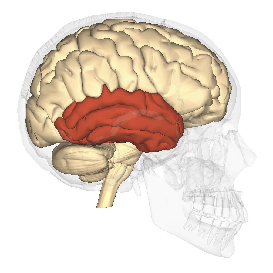
You actually have two temporal lobes, one for both hemispheres. Your temporal lobes are located near your ears, and they mainly function in auditory processing. The temporal lobe taps into other roles as well, such as visual memory, your ability to learn language, and emotion association.
Parietal Lobe
The parietal lobe is set between the occipital lobe and the frontal lobe.
The parietal lobe has a really big job of having to process the information that it receives within milliseconds. The somatosensory cortex is located inside of the parietal lobe, and is crucial for processing the information from touch. The somatosensory cortex allows us to pinpoint the exact locality of touch perception, and helps to distinguish pain levels and temperature.
The parietal lobe is important for our depth of perception and communicates with other parts of the brain in order to perform other tasks.
Occipital Lobe
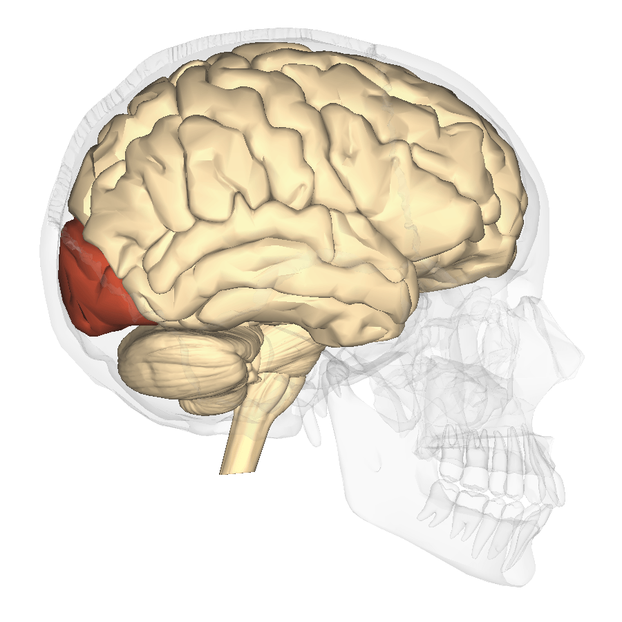
The occipital lobe sits in the back part of the cortex.
The main function of the occipital lobe is to process visual information from the eyes and relay that information. The occipital lobe has to very quickly process that information, so that we are able to respond accordingly.
Medulla
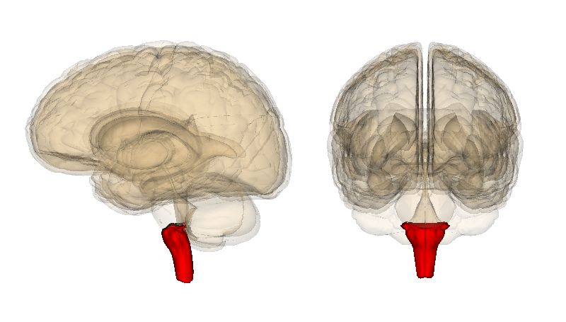
That is an overview of the cerebrum, now let’s look at the medulla, more officially known as the medulla oblongata. The medulla is a section of your brainstem, and remember, your brainstem is where your brain and spinal cord connect. The medulla is one of the three parts of the brainstem.
The medulla has immediate control over numerous autonomic nervous system responses, it aids in the control of particular regions of the body, and it also plays a role in motor functions and outward motion. The medulla is very important, just like every other part of our brain. The medulla helps control breathing, and it controls our heart rate.
The medulla functions involuntarily. The medulla responds to various heart functions by dilating the blood flow so that there is a higher or lower amount of oxygen circulation, it can help by responding in fight or flight situations by switching digestion off or on, causes us to cough or sneeze to get rid of unwanted particles that get into your nasal cavity, and also controls vomiting or swallowing in order to dispel anything that may bring harm to you.
Brain Stem
The fourth region of the brain that we will look at is the brain stem. The brain stem has three different parts: the medulla (which we just looked at), pons, and midbrain.
The pons sort of functions as a message headquarters for many other parts of the brain.
It aids in communication between brain sections, like the cerebrum and cerebellum. The pons is right above the medulla, and beneath the midbrain. So, it sits right in the middle. Many essential nerves stem from the pons. The vestibulocochlear nerve permits sound to proceed from our ears into our brain. The abducens nerve permits the eyes to move and look horizontally. The trigeminal nerve allows for us to experience feeling in the face, and the facial nerve allows for us to actually make facial expressions. The pons helps in the duties of the medulla, and it is linked to managing our sleep cycles.
Now, the midbrain is called the midbrain, because it sits between the forebrain and the hindbrain; but it’s important to note that out of the three parts of the brain stem it is not located in the middle. The midbrain functions, like the medulla, as this super message headquarters between the forebrain (or cerebral cortex) and the hindbrain (or the cerebellum and brainstem). The midbrain allows you to incorporate sensory data coming from your eyes and ears with data coming from your body’s muscle movements; helping it to adjust your movements based on important information.
All three parts of the brainstem function involuntarily.
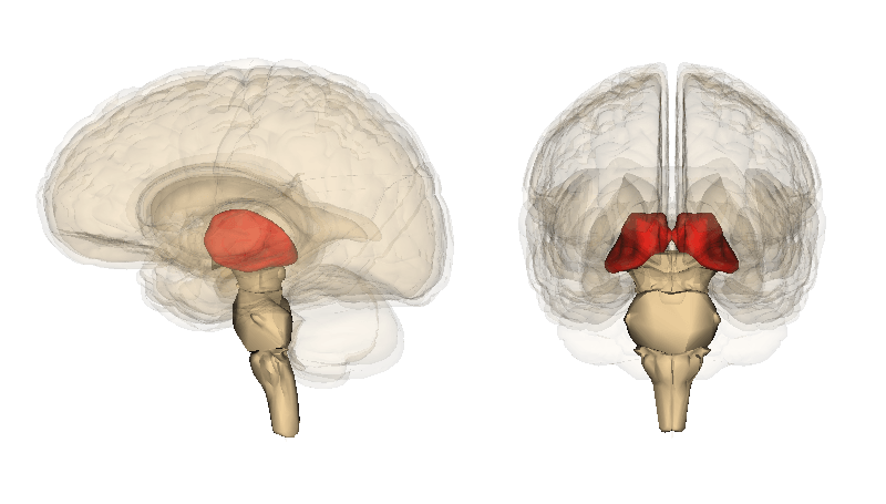
Thalamus
The thalamus is the fifth region of the brain. The thalamus sits right above the brainstem sandwiched between the midbrain and cerebral cortex.
The primary job of the thalamus is to inform the cerebral cortex of any sensory or motor stimuli. The thalamus also helps to control sleep, and aids in keeping us awake and alert. The thalamus, along with the hypothalamus, epithalamus, and subthalamus, is a part of the diencephalon, also sometimes called the interbrain. The diencephalon is the area where the vertebrate neural tube is located, which helps bring about the formation of the forebrain.
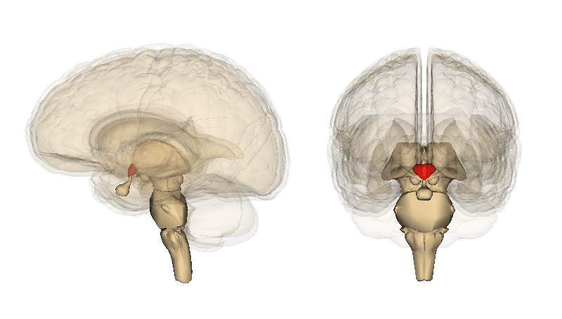
Hypothalamus
The sixth, and last, region of the brain is the hypothalamus. The hypothalamus is positioned below the thalamus and is the bottom part of the diencephalon.
One of the main functions of the hypothalamus is that it connects the nervous system to the endocrine system; which allows the hypothalamus to regulate our body’s temperature. The hypothalamus also aids in certain metabolic processes and helps to regulate the autonomic nervous system.
That was a closer look at the brain itself. The spinal cord is the other component that makes up the central nervous system. The spinal cord administers motor data coming from our brain to the appropriate parts of our body and helps to perform slight reflexes. It also administers sensory data directly from the peripheral nervous system to the brain. The spinal cord consists of 31 pairs of spinal nerves; because each of these nerves includes motor and sensory axons, they are referred to as mixed nerves.
So, that was the central nervous system. The second, main division of the nervous system is the peripheral nervous system.
Peripheral Nervous System
The peripheral nervous system is made up of nerves and what is called the ganglia.
Nerves are bundles of nerve fibers, also known as axons.
There are two types of nerves in the peripheral nervous system: spinal nerves and cranial nerves. There are 31 pairs of spinal nerves, and 12 pairs of cranial nerves. Ganglia refers to a cluster of neuron somas (or cell bodies). The individual cell body attaches to dendrites. The dendrites send information to neurons. The axons or nerve fibers then communicate that information to other neurons. It’s all a connected web. These nerves and ganglia are located around and outside of the brain and spinal cord.
The primary function of the peripheral nervous system is to link the central nervous system to the body’s organs and appendages, thus acting as the primary messenger between the brain, spinal cords, and everything else in the body. You can kind of think of the peripheral nervous system as the system that helps our brains and bodies to understand the world around us.
The peripheral nervous system has two subsystems: the somatic nervous system, and the autonomic nervous system.
Somatic Nervous System
The somatic nervous system is the part of the peripheral nervous system that is linked to voluntary bodily movements. It contains two parts, the sensory nervous system, as well as the somatosensory system, which is a part of the sensory nervous system.
The sensory nervous system is responsible for processing, as you might guess, sensory data. The most familiar sensory systems are the ones that control touch, taste, smell, hearing, and vision. The somatosensory system focuses on the conscious recognition of temperature, pain, touch, pressure, movement, position, and any sort of vibration. The somatosensory system communicates different sensations identified near the vicinity of the body via the spinal cord, brainstem, and up to the sensory cortex located in the parietal lobe. The sensory nervous system contains sensory neurons (or afferent neurons) and motor neurons (efferent neurons). Afferent neurons transmit stimuli to the central nervous system, as opposed to transmitting them from the CNS. Efferent neurons, in response to the afferent neurons, transmit signals from the central nervous system to the rest of the body. This transmission of stimuli through afferent neurons to the CNS, and then from the CNS to other parts of our body, allow for us to perform voluntary body movements as well as moderate involuntary reflex arcs.
Autonomic Nervous System
The autonomic nervous system is the other part of the peripheral nervous system that manages involuntary movements and functions—involuntary meaning that these movements happen without us be consciously in control of them happening. The autonomic nervous system also communicates information to our internal organs. There are two main divisions within the autonomic nervous system: the sympathetic nervous system, and the parasympathetic nervous system.
Sympathetic Nervous System
The sympathetic nervous system is the system that gets our body ready for the reaction that has been familiarized as the “fight or flight” response. When our body is being threatened or at risk for potential injury, this is the system that controls our response. The sympathetic nervous system causes our bodies to become keenly alert, speeds our heart rate, causes muscle contraction, and shuts off any function that is not essential for survival.
The overall functioning of the sympathetic nervous system is largely due to the neurotransmitter acetylcholine. This neurotransmitter binds to nicotinic receptors to trigger the release of norepinephrine which is produced by the adrenal gland to prepare our body and brain for action. Nicotinic receptors are the main receptors in the muscles for motor-neuron-to-muscle correspondence, that manage the contraction of muscles. The sympathetic nervous system originates in the thoracic and lumbar regions of the spinal cord.
Parasympathetic Nervous System
The parasympathetic nervous system, which is the other part of the autonomic nervous system, manages the body’s function when at rest. Rest and digest is the phrase given to reference the response of the parasympathetic nervous system. The parasympathetic nervous system works to conserve homeostasis within the body. It does the opposite of the sympathetic nervous system, and instead relaxes the body’s muscles, and slows our heart rate. Its job is to restore everything to a state of relaxation, and balance.
The parasympathetic nervous system increases bowel movements and urinary output. The neurotransmitter acetylcholine also plays a huge role in the parasympathetic nervous system by binding to muscarinic receptors, allowing the contraction of smooth muscles. This is what allows the parasympathetic to slow down the heart rate, regulate digestion, and relax muscles. The parasympathetic nervous system originates in the sacral portion of the spinal cord.
It’s important to note that both the central and peripheral nervous systems enlist the acetylcholine receptors, both nicotinic and muscarinic.
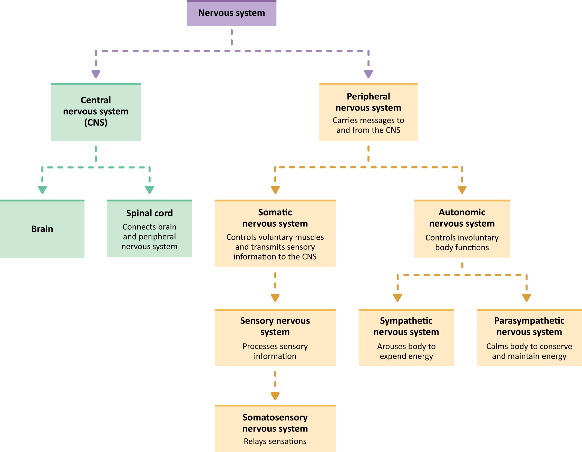
We just covered a lot of information. But, here is a very, very generalized diagram of the nervous system to help you remember the main parts.
You start with the nervous system itself. Then, you have the two main divisions: the central nervous system (which includes the brain and spinal cord), and the peripheral nervous system (which is made up of nerves and ganglia). The peripheral nervous system then has two subdivisions: the somatic nervous system (which controls voluntary movement), and the autonomic nervous system (which controls involuntary movement). Then, both the somatic and autonomic nervous systems contain two subdivisions. The somatic nervous system contains the sensory nervous system and the somatosensory system (which is a part of the sensory nervous system). And lastly, the autonomic nervous system contains the sympathetic nervous system (which controls our “fight or flight” response), and the parasympathetic nervous system which controls our “rest and digest” response).
If all of these systems seem pretty interconnected, the answer is yes.
I hope that this overview of the nervous system has been helpful to you!
What are the parts of the nervous system?
The nervous system has two main parts:
The nervous system transmits signals between the brain and the rest of the body, including internal organs. In this way, the nervous system’s activity controls the ability to move, breathe, see, think, and more. 1
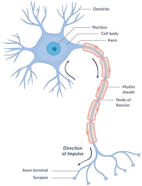
The basic unit of the nervous system is a nerve cell, or neuron. The human brain contains about 100 billion neurons. A neuron has a cell body, which includes the cell nucleus, and special extensions called axons (pronounced AK-sonz) and dendrites (pronounced DEN-drahytz). Bundles of axons, called nerves, are found throughout the body. Axons and dendrites allow neurons to communicate, even across long distances.
Different types of neurons control or perform different activities. For instance, motor neurons transmit messages from the brain to the muscles to generate movement. Sensory neurons detect light, sound, odor, taste, pressure, and heat and send messages about those things to the brain. Other parts of the nervous system control involuntary processes. These include keeping a regular heartbeat, releasing hormones like adrenaline, opening the pupil in response to light, and regulating the digestive system.
When a neuron sends a message to another neuron, it sends an electrical signal down the length of its axon. At the end of the axon, the electrical signal changes to a chemical signal. The axon then releases the chemical signal with chemical messengers called neurotransmitters (pronounced noor-oh-TRANS-mit-erz) into the synapse (pronounced SIN-aps)—the space between the end of an axon and the tip of a dendrite from another neuron. The neurotransmitters move the signal through the synapse to the neighboring dendrite, which converts the chemical signal back into an electrical signal. The electrical signal then travels through the neuron and goes through the same conversion processes as it moves to neighboring neurons.
The nervous system also includes non-neuron cells, called glia (pronounced GLEE-uh). Glia perform many important functions that keep the nervous system working properly. For example, glia:
The brain is made up of many networks of communicating neurons and glia. These networks allow different parts of the brain to “talk” to each other and work together to control body functions, emotions, thinking, behavior, and other activities. 1,2,3
What You Need to Know About the Nervous System
Rod Brouhard is an emergency medical technician paramedic (EMT-P), journalist, educator, and advocate for emergency medical service providers and patients.
Nicholas R. Metrus, MD, is a board-certified neurologist and neuro-oncologist. He currently serves at the Glasser Brain Tumor Center in Summit, New Jersey.
The nervous system is an organ system that handles communication in the body. There are four types of nerve cells in the nervous system: sensory nerves, motor nerves, autonomic nerves and inter-neurons (neuron is just a fancy word for nerve cell).
You can divide up all the nerves in the body into roughly two parts: the central nervous system and the peripheral nervous system.
Central Nervous System (CNS)
The central nervous system contains two organs—the brain and the spinal cord. It has all four types of nerve cells and is the only place you can find inter-neurons. The central nervous system is insulated from the outside world pretty well. It never even touches blood. It gets its nutrients from cerebrospinal fluid, a clear liquid that bathes the brain and spinal cord.
Both organs are covered with three layers of membranes called the meninges. CITE The meninges and cerebrospinal fluid cushion the brain to keep it from being injured by a knock on the noggin. It’s possible to get an infection from viruses or bacteria in the meninges called meningitis. It’s also possible to have bleeding either between the meninges and the skull (called an epidural hematoma) or in between the layers of the meninges (called a subdural hematoma). Any bleeding or infection inside the skull can put pressure on the brain and cause it to malfunction.
The central nervous system is like the guts of your computer. It’s in there with millions of connections moving little impulses around from circuit to circuit (nerve to nerve), calculating and thinking. Your brain makes all the calculations and stores information. Your spinal cord is like a cable with lots of individual wires running to all different parts of the brain.
But the computer brain inside your laptop, like the brain inside your head, is useless all by itself. You have to be able to tell your computer what you need and see or hear what your computer is trying to tell you. You need some sort of input and output devices. Your computer uses a mouse, a touchscreen or a keyboard to sense what you want it to do. It uses a screen and speakers to react.
Your body works very similarly. You have sensory organs to send information to the brain—eyes, ears, nose, tongue, and skin. To react, you have muscles that make you walk, talk, focus, wink, stick your tongue out—whatever. Your input/output devices are part of your peripheral nervous system.
Peripheral Nervous System (PNS)
The peripheral nervous system is everything connected to the central nervous system. It has motor nerves, sensory nerves, and autonomic nerves. Autonomic nerves act automatically, which is a way to remember them. They are the nerves that regulate our bodies. They are the body’s version of a thermostat, a clock, and a smoke alarm. They work in the background to keep us on track and healthy, but they don’t take up brain power or need to be controlled.
Autonomic nerves are loosely split into either sympathetic or parasympathetic nerves.
Think of the sympathetic nerves as the body’s accelerator, and parasympathetic nerves as the brake pedal. Your body is always stimulating both the parasympathetic side and the sympathetic side at the same time—just like my grandmother used to drive, with a foot on each pedal.
Motor nerves start from the central nervous system and go out toward the far reaches of the body. They’re called motor nerves because they always end in muscles. If you think about it, the only signals your brain sends to the outside world consist of making things move. Walking, talking, fighting, running, or singing all take muscles.
A Word About the Spinal Cord
The spinal cord is the connection between the central nervous system and the peripheral. It is technically part of the CNS, but it is how most of the motor and sensory nerves get to the brain. Inside the spinal cord are some of those inter-neurons mentioned above. In the brain, inter-neurons are like the microscopic switches in a computer chip, helping to make calculations and do the heavy thinking.
In the spinal cord, inter-neurons have a different function. Here they act like a planned short circuit, letting us react to some things faster than we could if the signal had to travel all the way to the brain and back. Inter-neurons in the spinal cord are responsible for reflexes—the reason you jerk back when you touch a hot pan before you even realize what happened.
Sending Signals
Nerves carry messages via signals called impulses. Like a computer the signal is binary; it’s either on or off. A single nerve cell can’t send a weaker signal or a stronger signal. It can change frequency—ten impulses per second, for example, or thirty—but each impulse is exactly the same.
Impulses travel along a nerve in exactly the same way as muscle cells contract, through chemistry. Nerve cells use ionized minerals (salts like calcium, potassium, and sodium) to propel the impulse along. I won’t get too deep into the physiology, but the body needs a proper balance of all three of these minerals for the process to work correctly. Too much or too little of any of these and neither muscles nor nerves will function properly.
Nerve cells can be pretty long, but it still takes several to reach from the tip of your finger to your spinal cord. The cells don’t touch each other. Instead, the impulse is chemically sent (transmitted) from one nerve cell to the next using substances known as neurotransmitters.
Adding neurotransmitters to the bloodstream can cause nerves to send signals. For example, many of the sympathetic nerve cells mentioned above (the fight-or-flight cells) react to a neurotransmitter called adrenaline, which is released into the bloodstream from the adrenal glands when we get scared, stressed, or startled.
A Word From Verywell
If you have a solid grasp of how the nervous system works, it’s a small leap to understanding why certain substances or medications affect us the way they do. It’s also easier to understand how strokes or concussions affect the brain.
The body is a dynamic collection of chemicals constantly interacting. The nervous system is the most basic of those interactions. This is the foundation for understanding physiology as a whole.
National Institute of Neurological Disorders and Stroke. Meningitis and encephalitis fact sheet.
Nervous system
Author: Jana Vasković MD • Reviewer: Nicola McLaren MSc
Last reviewed: August 15, 2022
Reading time: 20 minutes
The nervous system is a network of neurons whose main feature is to generate, modulate and transmit information between all the different parts of the human body. This property enables many important functions of the nervous system, such as regulation of vital body functions (heartbeat, breathing, digestion), sensation and body movements. Ultimately, the nervous system structures preside over everything that makes us human; our consciousness, cognition, behaviour and memories.
The nervous system consists of two divisions;
Understanding the nervous system requires knowledge of its various parts, so in this article you will learn about the nervous system breakdown and all its various divisions.
Cells of the nervous system
Two basic types of cells are present in the nervous system;
Neurons
Neurons, or nerve cell, are the main structural and functional units of the nervous system. Every neuron consists of a body (soma) and a number of processes (neurites). The nerve cell body contains the cellular organelles and is where neural impulses (action potentials) are generated. The processes stem from the body, they connect neurons with each other and with other body cells, enabling the flow of neural impulses. There are two types of neural processes that differ in structure and function;
Every neuron has a single axon, while the number of dendrites varies. Based on that number, there are four structural types of neurons; multipolar, bipolar, pseudounipolar and unipolar.
Learn more about the neurons in our study unit:
How do neurons function?
The morphology of neurons makes them highly specialized to work with neural impulses; they generate, receive and send these impulses onto other neurons and non-neural tissues.
There are two types of neurons, named according to whether they send an electrical signal towards or away from the CNS;
The site where an axon connects to another cell to pass the neural impulse is called a synapse. The synapse doesn’t connect to the next cell directly. Instead, the impulse triggers the release of chemicals called neurotransmitters from the very end of an axon. These neurotransmitters bind to the effector cell’s membrane, causing biochemical events to occur within that cell according to the orders sent by the CNS.
Ready to reinforce your knowledge about the neurons? Try out our quiz below:
Glial cells
Glial cells, also called neuroglia or simply glia, are smaller non-excitatory cells that act to support neurons. They do not propagate action potentials. Instead, they myelinate neurons, maintain homeostatic balance, provide structural support, protection and nutrition for neurons throughout the nervous system.
This set of functions is provided for by four different types of glial cells;
Most axons are wrapped by a white insulating substance called the myelin sheath, produced by oligodendrocytes and Schwann cells. Myelin encloses an axon segmentally, leaving unmyelinated gaps between the segments called the nodes of Ranvier. The neural impulses propagate through the Ranvier nodes only, skipping the myelin sheath. This significantly increases the speed of neural impulse propagation.
White and gray matter
The white color of myelinated axons is distinguished from the gray colored neuronal bodies and dendrites. Based on this, nervous tissue is divided into white matter and gray matter, both of which has a specific distribution;
Master the histology of nervous tissue with our customizable quiz: We got you covered with neurons, nerves and ganglia!
Nervous system divisions
So nervous tissue, comprised of neurons and neuroglia, forms our nervous organs (e.g. the brain, nerves). These organs unite according to their common function, forming the evolutionary perfection that is our nervous system.
The nervous system (NS) is structurally broken down into two divisions;
Functionally, the PNS is further subdivided into two functional divisions;
They say that the nervous system is one of the hardest anatomy topic. But you’re in luck, as we’ve got a learning strategy for you to master neuroanatomy in a lot shorter time than you though you’ll need. Check out our quizzes and more for the nervous system anatomy practice!
Although divided structurally into central and peripheral parts, the nervous system divisions are actually interconnected with each other. Axon bundles pass impulses between the brain and spinal cord. These bundles within the CNS are called afferent and efferent neural pathways or tracts. Axons that extend from the CNS to connect with peripheral tissues belong to the PNS. Axons bundles within the PNS are called afferent and efferent peripheral nerves.
Central nervous system
The central nervous system (CNS) consists of the brain and spinal cord. These are found housed within the skull and vertebral column respectively.
The brain is made of four parts; cerebrum, diencephalon, cerebellum and brainstem. Together these parts process the incoming information from peripheral tissues and generate commands; telling the tissues how to respond and function. These commands tackle the most complex voluntary and involuntary human body functions, from breathing to thinking.
The spinal cord continues from the brainstem. It also has the ability to generate commands but for involuntary processes only, i.e. reflexes. However, its main function is to pass information between the CNS and periphery.
Learn more about the CNS anatomy here:
Peripheral nervous system
The PNS consists of 12 pairs of cranial nerves, 31 pairs of spinal nerves and a number of small neuronal clusters throughout the body called ganglia.
Peripheral nerves can be sensory (afferent), motor (efferent) or mixed (both). Depending on what structures they innervate, peripheral nerves can have the following modalities;
Cranial nerves
Cranial nerves are peripheral nerves that emerge from the cranial nerve nuclei of the brainstem and spinal cord. They innervate the head and neck. Cranial nerves are numbered one to twelve according to their order of exit through the skull fissures. Namely, they are: olfactory nerve (CN I), optic nerve (CN II), oculomotor nerve (CN III), trochlear nerve (CN IV), trigeminal nerve (CN V), abducens nerve (VI), facial nerve (VII), vestibulocochlear nerve (VIII), glossopharyngeal nerve (IX), vagus nerve (X), accessory nerve (XI), and hypoglossal nerve (XII). These nerves are motor (III, IV, VI, XI, and XII), sensory (I, II and VIII) or mixed (V, VII, IX, and X).
Among many strategies for learning cranial nerves anatomy, our experts have determined that one of the most efficient is through interactive learning. Check out Kenhub’s interactive cranial nerves quizzes and labeling exercises to cut your studying time in half.
Jump right into our cranial nerves quiz in multiple difficulty levels:
Or learn more about the cranial nerves in this study unit.
Spinal nerves
Spinal nervesemerge from the segments of the spinal cord. They are numbered according to their specific segment of origin. Hence, the 31 pairs of spinal nerves are divided into 8 cervical pairs, 12 thoracic pairs, 5 lumbar pairs, 5 sacral pairs, and 1 coccygeal spinal nerve. All spinal nerves are mixed, containing both sensory and motor fibers.
Spinal nerves innervate the entire body, with the exception of the head. They do so by either directly synapsing with their target organs or by interlacing with each other and forming plexuses. There are four major plexuses that supply the body regions;
Want to learn more about the spinal nerves and plexuses? Check out our resources.
Ganglia
Ganglia (sing. ganglion) are clusters of neuronal cell bodies outside of the CNS, meaning that they are the PNS equivalents to subcortical nuclei of the CNS. Ganglia can be sensory or visceral motor (autonomic) and their distribution in the body is clearly defined.
Dorsal root ganglia are clusters of sensory nerve cell bodies located adjacent to the spinal cord, They are a component of the posterior root of a spinal nerve.
Autonomic ganglia are either sympathetic or parasympathetic. Sympathetic ganglia are found in the thorax and abdomen, grouped into paravertebral and prevertebral ganglia. Paravertebral ganglia lie on either side of vertebral column (para- means beside), comprising two ganglionic chains that extend from the base of the skull to the coccyx, called sympathetic trunks. Prevertebral ganglia (collateral ganglia, preaortic ganglia) are found anterior to the vertebral column (pre- means in front of), closer to their target organ. They are further grouped according to which branch of abdominal aorta they surround; celiac, aorticorenal, superior and inferior mesenteric ganglia.
Parasympathetic ganglia are found in the head and pelvis. Ganglia in the head are associated with relevant cranial nerves and are the ciliary, pterygopalatine, otic and submandibular ganglia. Pelvic ganglia lie close to the reproductive organs comprising autonomic plexuses for innervation of pelvic viscera, such as prostatic and uterovaginal plexuses.
Find everything about ganglia needed for your neuroanatomy exam here.
Somatic nervous system
The somatic nervous system is the voluntary component of the peripheral nervous system. It consists of all the fibers within cranial and spinal nerves that enable us to perform voluntary body movements (efferent nerves) and feel sensation from the skin, muscles and joints (afferent nerves). Somatic sensation relates to touch, pressure, vibration, pain, temperature, stretch and position sense from these three types of structures.
Sensation from the glands, smooth and cardiac muscles is conveyed by the autonomic nerves.
Nervous System
The nervous system consists of the brain, spinal cord, sensory organs, and all of the nerves that connect these organs with the rest of the body. Together, these organs are responsible for the control of the body and communication among its parts. The brain and spinal cord form the control center known as the central nervous system (CNS), where information is evaluated and decisions made. The sensory nerves and sense organs of the peripheral nervous system (PNS) monitor conditions inside and outside of the body and send this information to the CNS. Efferent nerves in the PNS carry signals from the control center to the muscles, glands, and organs to regulate their functions. Continue Scrolling To Read More Below.
Additional Resources
Anatomy Explorer
Change Current View Angle
Toggle Anatomy System
Switch Gender
Anatomy Term
Displayed on other page
Nervous System Anatomy
Nervous Tissue
The majority of the nervous system is tissue made up of two classes of cells: neurons and neuroglia.
Neurons
Neurons, also known as nerve cells, communicate within the body by transmitting electrochemical signals. Neurons look quite different from other cells in the body due to the many long cellular processes that extend from their central cell body. The cell body is the roughly round part of a neuron that contains the nucleus, mitochondria, and most of the cellular organelles. Small tree-like structures called dendrites extend from the cell body to pick up stimuli from the environment, other neurons, or sensory receptor cells. Long transmitting processes called axons extend from the cell body to send signals onward to other neurons or effector cells in the body.
There are 3 basic classes of neurons: afferent neurons, efferent neurons, and interneurons.
Neuroglia
Neuroglia, also known as glial cells, act as the “helper” cells of the nervous system. Each neuron in the body is surrounded by anywhere from 6 to 60 neuroglia that protect, feed, and insulate the neuron. Because neurons are extremely specialized cells that are essential to body function and almost never reproduce, neuroglia are vital to maintaining a functional nervous system.
Brain
The brain, a soft, wrinkled organ that weighs about 3 pounds, is located inside the cranial cavity, where the bones of the skull surround and protect it. The approximately 100 billion neurons of the brain form the main control center of the body. The brain and spinal cord together form the central nervous system (CNS), where information is processed and responses originate. The brain, the seat of higher mental functions such as consciousness, memory, planning, and voluntary actions, also controls lower body functions such as the maintenance of respiration, heart rate, blood pressure, and digestion.
Spinal Cord
The spinal cord is a long, thin mass of bundled neurons that carries information through the vertebral cavity of the spine beginning at the medulla oblongata of the brain on its superior end and continuing inferiorly to the lumbar region of the spine. In the lumbar region, the spinal cord separates into a bundle of individual nerves called the cauda equina (due to its resemblance to a horse’s tail) that continues inferiorly to the sacrum and coccyx. The white matter of the spinal cord functions as the main conduit of nerve signals to the body from the brain. The grey matter of the spinal cord integrates reflexes to stimuli.
Nerves
Nerves are bundles of axons in the peripheral nervous system (PNS) that act as information highways to carry signals between the brain and spinal cord and the rest of the body. Each axon is wrapped in a connective tissue sheath called the endoneurium. Individual axons of the nerve are bundled into groups of axons called fascicles, wrapped in a sheath of connective tissue called the perineurium. Finally, many fascicles are wrapped together in another layer of connective tissue called the epineurium to form a whole nerve. The wrapping of nerves with connective tissue helps to protect the axons and to increase the speed of their communication within the body.
Meninges
The meninges are the protective coverings of the central nervous system (CNS). They consist of three layers: the dura mater, arachnoid mater, and pia mater.
Cerebrospinal Fluid
The space surrounding the organs of the CNS is filled with a clear fluid known as cerebrospinal fluid (CSF). CSF is formed from blood plasma by special structures called choroid plexuses. The choroid plexuses contain many capillaries lined with epithelial tissue that filters blood plasma and allows the filtered fluid to enter the space around the brain.
Newly created CSF flows through the inside of the brain in hollow spaces called ventricles and through a small cavity in the middle of the spinal cord called the central canal. CSF also flows through the subarachnoid space around the outside of the brain and spinal cord. CSF is constantly produced at the choroid plexuses and is reabsorbed into the bloodstream at structures called arachnoid villi.
Cerebrospinal fluid provides several vital functions to the central nervous system:
Sense Organs
All of the bodies’ many sense organs are components of the nervous system. What are known as the special senses—vision, taste, smell, hearing, and balance—are all detected by specialized organs such as the eyes, taste buds, and olfactory epithelium. Sensory receptors for the general senses like touch, temperature, and pain are found throughout most of the body. All of the sensory receptors of the body are connected to afferent neurons that carry their sensory information to the CNS to be processed and integrated.
Nervous System Physiology
Functions of the Nervous System
The nervous system has 3 main functions: sensory, integration, and motor.
Divisions of the Nervous System
Central Nervous System
The brain and spinal cord together form the central nervous system, or CNS. The CNS acts as the control center of the body by providing its processing, memory, and regulation systems. The CNS takes in all of the conscious and subconscious sensory information from the body’s sensory receptors to stay aware of the body’s internal and external conditions. Using this sensory information, it makes decisions about both conscious and subconscious actions to take to maintain the body’s homeostasis and ensure its survival. The CNS is also responsible for the higher functions of the nervous system such as language, creativity, expression, emotions, and personality. The brain is the seat of consciousness and determines who we are as individuals.
Peripheral Nervous System
The peripheral nervous system (PNS) includes all of the parts of the nervous system outside of the brain and spinal cord. These parts include all of the cranial and spinal nerves, ganglia, and sensory receptors.
Somatic Nervous System
The somatic nervous system (SNS) is a division of the PNS that includes all of the voluntary efferent neurons. The SNS is the only consciously controlled part of the PNS and is responsible for stimulating skeletal muscles in the body.
Autonomic Nervous System
The autonomic nervous system (ANS) is a division of the PNS that includes all of the involuntary efferent neurons. The ANS controls subconscious effectors such as visceral muscle tissue, cardiac muscle tissue, and glandular tissue.
There are 2 divisions of the autonomic nervous system in the body: the sympathetic and parasympathetic divisions.
Enteric Nervous System
The enteric nervous system (ENS) is the division of the ANS that is responsible for regulating digestion and the function of the digestive organs. The ENS receives signals from the central nervous system through both the sympathetic and parasympathetic divisions of the autonomic nervous system to help regulate its functions. However, the ENS mostly works independently of the CNS and continues to function without any outside input. For this reason, the ENS is often called the “brain of the gut” or the body’s “second brain.” The ENS is an immense system—almost as many neurons exist in the ENS as in the spinal cord.
Action Potentials
Neurons function through the generation and propagation of electrochemical signals known as action potentials (APs). An AP is created by the movement of sodium and potassium ions through the membrane of neurons. (See Water and Electrolytes.)
Synapses
A synapse is the junction between a neuron and another cell. Synapses may form between 2 neurons or between a neuron and an effector cell. There are two types of synapses found in the body: chemical synapses and electrical synapses.
Myelination
The axons of many neurons are covered by a coating of insulation known as myelin to increase the speed of nerve conduction throughout the body. Myelin is formed by 2 types of glial cells: Schwann cells in the PNS and oligodendrocytes in the CNS. In both cases, the glial cells wrap their plasma membrane around the axon many times to form a thick covering of lipids. The development of these myelin sheaths is known as myelination.
Myelination speeds up the movement of APs in the axon by reducing the number of APs that must form for a signal to reach the end of an axon. The myelination process begins speeding up nerve conduction in fetal development and continues into early adulthood. Myelinated axons appear white due to the presence of lipids and form the white matter of the inner brain and outer spinal cord. White matter is specialized for carrying information quickly through the brain and spinal cord. The gray matter of the brain and spinal cord are the unmyelinated integration centers where information is processed.
Reflexes
Reflexes are fast, involuntary responses to stimuli. The most well known reflex is the patellar reflex, which is checked when a physicians taps on a patient’s knee during a physical examination. Reflexes are integrated in the gray matter of the spinal cord or in the brain stem. Reflexes allow the body to respond to stimuli very quickly by sending responses to effectors before the nerve signals reach the conscious parts of the brain. This explains why people will often pull their hands away from a hot object before they realize they are in pain.
Functions of the Cranial Nerves
Each of the 12 cranial nerves has a specific function within the nervous system.
Sensory Physiology
All sensory receptors can be classified by their structure and by the type of stimulus that they detect. Structurally, there are 3 classes of sensory receptors: free nerve endings, encapsulated nerve endings, and specialized cells. Free nerve endings are simply free dendrites at the end of a neuron that extend into a tissue. Pain, heat, and cold are all sensed through free nerve endings. An encapsulated nerve ending is a free nerve ending wrapped in a round capsule of connective tissue. When the capsule is deformed by touch or pressure, the neuron is stimulated to send signals to the CNS. Specialized cells detect stimuli from the 5 special senses: vision, hearing, balance, smell, and taste. Each of the special senses has its own unique sensory cells—such as rods and cones in the retina to detect light for the sense of vision.
Functionally, there are 6 major classes of receptors: mechanoreceptors, nociceptors, photoreceptors, chemoreceptors, osmoreceptors, and thermoreceptors.



:max_bytes(150000):strip_icc()/Rod-Brouhard-EMT-P_1000-85058e33388640c0a9cc5e20427cfe90.jpg)
:max_bytes(150000):strip_icc()/Nicholas-Metrus_1000-41893b3fb9e84f3e99cf0f18b2464fdb.jpg)
:max_bytes(150000):strip_icc()/brain_neuron_activity-57d880483df78c5833a884c6.jpg)
:background_color(FFFFFF):format(jpeg)

